Home
/ Diagram Of Animal Cell Under Light Microscope : Animal Cell Under Microscope Structure And Anatomy / Animal cells are not only tiny but they are also colorless and translucent.
Diagram Of Animal Cell Under Light Microscope : Animal Cell Under Microscope Structure And Anatomy / Animal cells are not only tiny but they are also colorless and translucent.
Diagram Of Animal Cell Under Light Microscope : Animal Cell Under Microscope Structure And Anatomy / Animal cells are not only tiny but they are also colorless and translucent.. The animal cell is made up of. Cells under microscope foto sin derechos de autor. 3) a student viewing a specimen under low power of a compound light microscope switched to high 17) microscopic examination of an animal cell reveals the presence of a plasma membrane but no cell. You should not look through the microscope to do this. Plant cells have cell walls, one large vacuole per cell, and chloroplasts, while animal cells will have a cell membrane only.
With light microscopy i can simply scrape some cells from my cheek smear them on a slide and look at them. Observing a wide range of biological processes and animal cell under light microscope is easier due to advances in microscopic techniques. What invention made it possibe for people to study cells? Animal cells are not only tiny but they are also colorless and translucent. However, it is useful to know roughly where they are and what they do.
A Typical Animal Cell As Seen In An Electron Microscope Medical Ima from image.slidesharecdn.com Use the diagram to show how the microscope works (i.e. This is a diagram of a typical plant cell. It also has a very high resolving power. Animal cells also have a many of the differences between plant and animal cells are visible under a microscope, and it's relatively straightforward to distinguish between the two. Some features common to animal cells. Observing a wide range of biological processes and animal cell under light microscope is easier due to advances in microscopic techniques. Preparing onion cell slides is a useful way to observe simple plant cells under the light microscope. Made the first compound microscope and observed a slice of a cork oak tree.
To use a light microscope to examine animal or plant cells.
Gcse biology microscope drawing and measuring cell size edexcel 9 1. To use a light microscope to examine animal or plant cells. Albeit the detail will be minimal without a contrast mechanism or staining or such. Use the diagram to show how the microscope works (i.e. The role and function of the plasma membrane; Preparing onion cell slides is a useful way to observe simple plant cells under the light microscope. Animal cells are not only tiny but they are also colorless and translucent. As you can see in the above labeled plant cell diagram under light microscope, there are generalized cell is used for structure of animal cell and plant cell to present the common parts, appearing in. Observe the onion skin under low power of the microscope and then under high power. Image:animal cell seen under electron microscope. Cell is a tiny structure and functional unit of a living organism containing various parts known as organelles. Students will observe onion cells under a microscope. Image:plant cell seen under electron microscope.
Observing a wide range of biological processes and animal cell under light microscope is easier due to advances in microscopic techniques. Examining animal cells under the microscope. With light microscopy i can simply scrape some cells from my cheek smear them on a slide and look at them. Simple diagrams of a light microscope. That cells can be of different shapes and sizes.
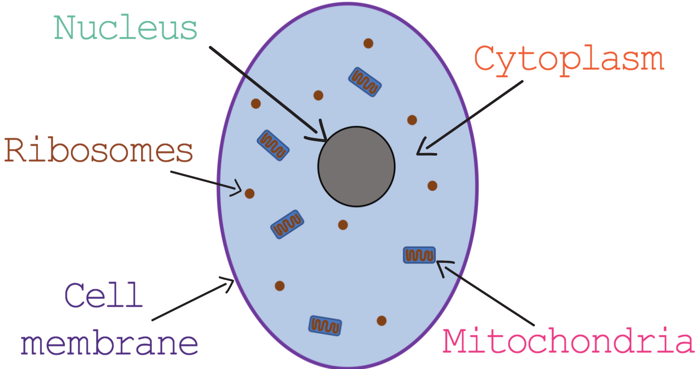
E Ig Hm 1 1 Elevise from www.elevise.co.uk A scale bar has been marked on the drawing, allowing the size of a cell to be estimated. 3) a student viewing a specimen under low power of a compound light microscope switched to high 17) microscopic examination of an animal cell reveals the presence of a plasma membrane but no cell. If a plant cell and an animal cell are observed under a microscope, what are the characteristics of the cells. According to your observations in this lab and your book and notes, create a venn diagram to illustrate the structural similarities and differences between plant and animal cells. This is a diagram of a typical plant cell. What was once unseeable can now be seen, touched, and eaten!cut. This shows a generalized animal cell under a light microscope. Cell structure teaching resources the science teacher, organelles biology for majors i, 11 different types of cells in the human body, class test, chronic inflammation under the microscope learn share.
We say cells are microscopic because they can only be seen under a microscope.
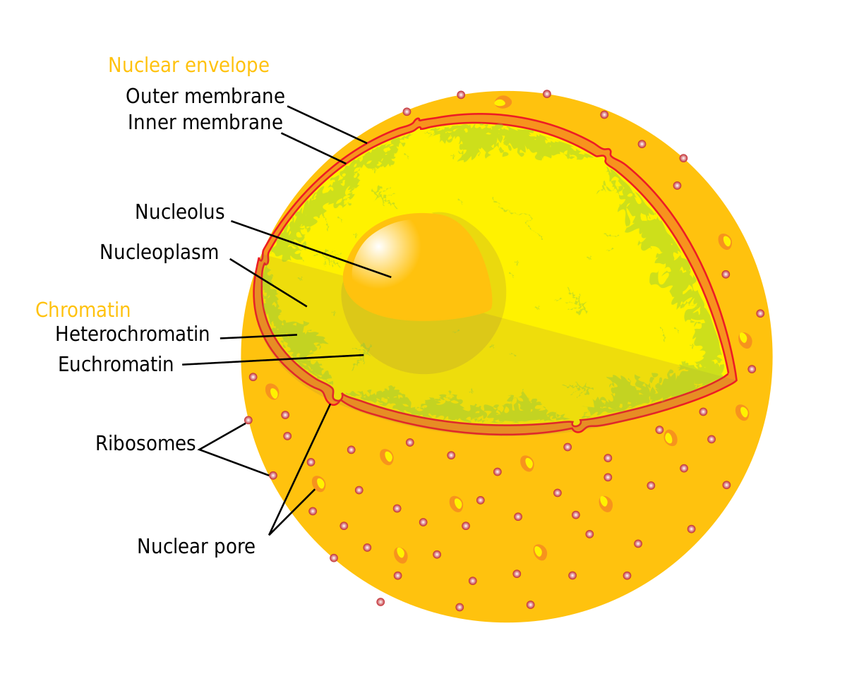
Cell is a tiny structure and functional unit of a living organism containing various parts known as organelles. It also has a very high resolving power. Draw your own light microscope and label the parts. Image:plant cell seen under electron microscope. Plant cell science diagram clipart set includes: Cytoplasm, ribosomes, rough endoplasmic reticulum; Describe and compare the structure of a plant cell with an animal cell, as seen under a light microscope, limited to cell wall, nucleus, cytoplasm, chloroplasts, vacuoles and location of the cell membrane. A generalised animal cell as observed under an electron microscope. Line diagram of a general animal cell. We use microscope comprehensively in microbiology, mineralogy, cell biology, biotechnology, nano physics, microelectronics, pharmacology, and forensics. Light microscopes using visible light and lenses to form a magnified image of the object under investigation e.g. Image:animal cell seen under electron microscope. Study the two diagrams of plant and animal cells below.
Plant cell science diagram clipart set includes: If a plant cell and an animal cell are observed under a microscope, what are the characteristics of the cells. Some features common to animal cells. Cell is a tiny structure and functional unit of a living organism containing various parts known as organelles. Physiology is the study of the functions of the body at the.
Nucleolus Wikipedia from upload.wikimedia.org Preparing onion cell slides is a useful way to observe simple plant cells under the light microscope. With light microscopy i can simply scrape some cells from my cheek smear them on a slide and look at them. Cytoplasm, ribosomes, rough endoplasmic reticulum; Plant and animal cells lab objectives:. The animal cell is made up of. However, it is useful to know roughly where they are and what they do. This is a diagram of a typical plant cell. Since animal cells lack a rigid cell wall it allows them to develop a great diversity of cell types, tissues, and note:
Cell structure teaching resources the science teacher, organelles biology for majors i, 11 different types of cells in the human body, class test, chronic inflammation under the microscope learn share.
Physiology is the study of the functions of the body at the. Students will observe onion cells under a microscope. The animal cell is made up of. See how a generalized structure of an animal cell and plant cell look with labeled diagrams. Light microscopes are used in biology classes in schools and colleges as well as in professional scientific environments such as government laboratories and biotechnology companies. Some features common to animal cells. 3) a student viewing a specimen under low power of a compound light microscope switched to high 17) microscopic examination of an animal cell reveals the presence of a plasma membrane but no cell. Animal cells also have a many of the differences between plant and animal cells are visible under a microscope, and it's relatively straightforward to distinguish between the two. Image:animal cell seen under electron microscope. Diagram of animal cell, created with biorender.com. Smooth endoplasmic reticulum, mitochondria, golgi bodies, lysosomes. Since animal cells lack a rigid cell wall it allows them to develop a great diversity of cell types, tissues, and note: Made the first compound microscope and observed a slice of a cork oak tree.
Share :
Post a Comment
for "Diagram Of Animal Cell Under Light Microscope : Animal Cell Under Microscope Structure And Anatomy / Animal cells are not only tiny but they are also colorless and translucent."


Post a Comment for "Diagram Of Animal Cell Under Light Microscope : Animal Cell Under Microscope Structure And Anatomy / Animal cells are not only tiny but they are also colorless and translucent."